
A biopsy of blastomeres is carried out on day 3 of embyo development for all embryos obtained in the IVF cycle that have 5 or more blastomeres with a degree of fragmentation not higher than 25%.
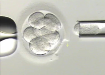
A biopsy of a blastomere on day 3 of the development
The ADVANTAGES are:
- The method allows the analysis not only of meiotic errors of the paternal and the maternal genome but also of mitotic ones;
- Enough time for genetic diagnosis;
- An embryo transfer in the same cycle;
- The biopsy can be repeated.
- Earlier studies claimed there was no harm for an embryo (Hardy et al., 1989), yet the modern ones speak about embryo damage (Mastenbroek et al, 2007, Scott et al, 2012);
- A decrease in the potential of implantation (Cohen et al, 2010);
- A high level of chromosomal mosaicism of pre-implantation embryos; a possibility that a healthy embryo can be diagnosed with a pathology, which nullifies the efficacy of diagnosis.
- A decrease in the number of ICM and trophectoderm cells;
- A decrease in HCG-producing activity of a trophoblast when implanting embryos that have undergone a biopsy (Yeon Jean Cho et al, 2011).
- A change in the expression of 36 genes;
- Hypomielinization of nerve fibers in the brain;
- An increased risk of developing neurodegenerative diseases.
This is educational material from the portal’s archive. It does not replace medical advice. Discuss tests, medicines and treatment decisions with your clinician.